CASE20230818_002
Rota-Shock Treat Severe Calcification Case
By Songbai Deng, Guozhu Chen
Presenter
Guozhu Chen
Authors
Songbai Deng1, Guozhu Chen1
Affiliation
Department of Cardiology, The Second Affiliated Hospital of Chongqing Medical University., China1,
View Study Report
CASE20230818_002
Complex PCI - Calcified Lesion
Rota-Shock Treat Severe Calcification Case
Songbai Deng1, Guozhu Chen1
Department of Cardiology, The Second Affiliated Hospital of Chongqing Medical University., China1,
Clinical Information
Relevant Clinical History and Physical Exam
A male, 77 years old with Exertional chest pain for 4 months admitted to our department.CV history: No DM/HBP, No smoking. Pre-Angiograms finished three months ago: LAD: severe calcification, proximal LAD with stenosis of about 90%, LCX: stenosis of about 70-80%, RCA severe calcification, subtotal occlusion at the proximal segment; LAD to RCA distal collateral. Aspirin enteric-coated tablets (Bayaspirin Enteric-coated) 100mg/d, Clopidogrel (Plavix) 75mg/d, and Atorvastatin 20mg/d.


Relevant Test Results Prior to Catheterization
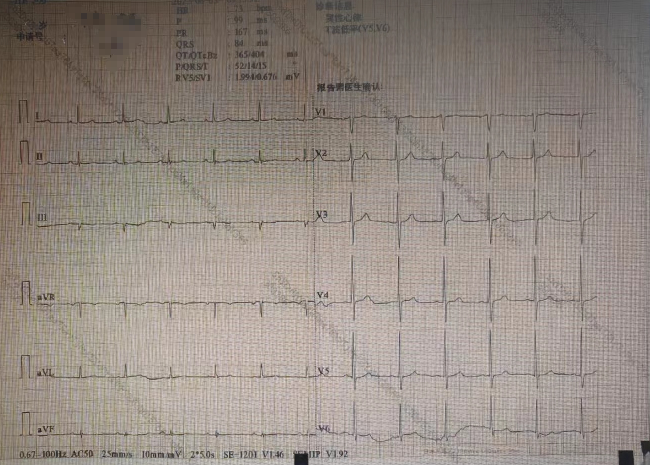
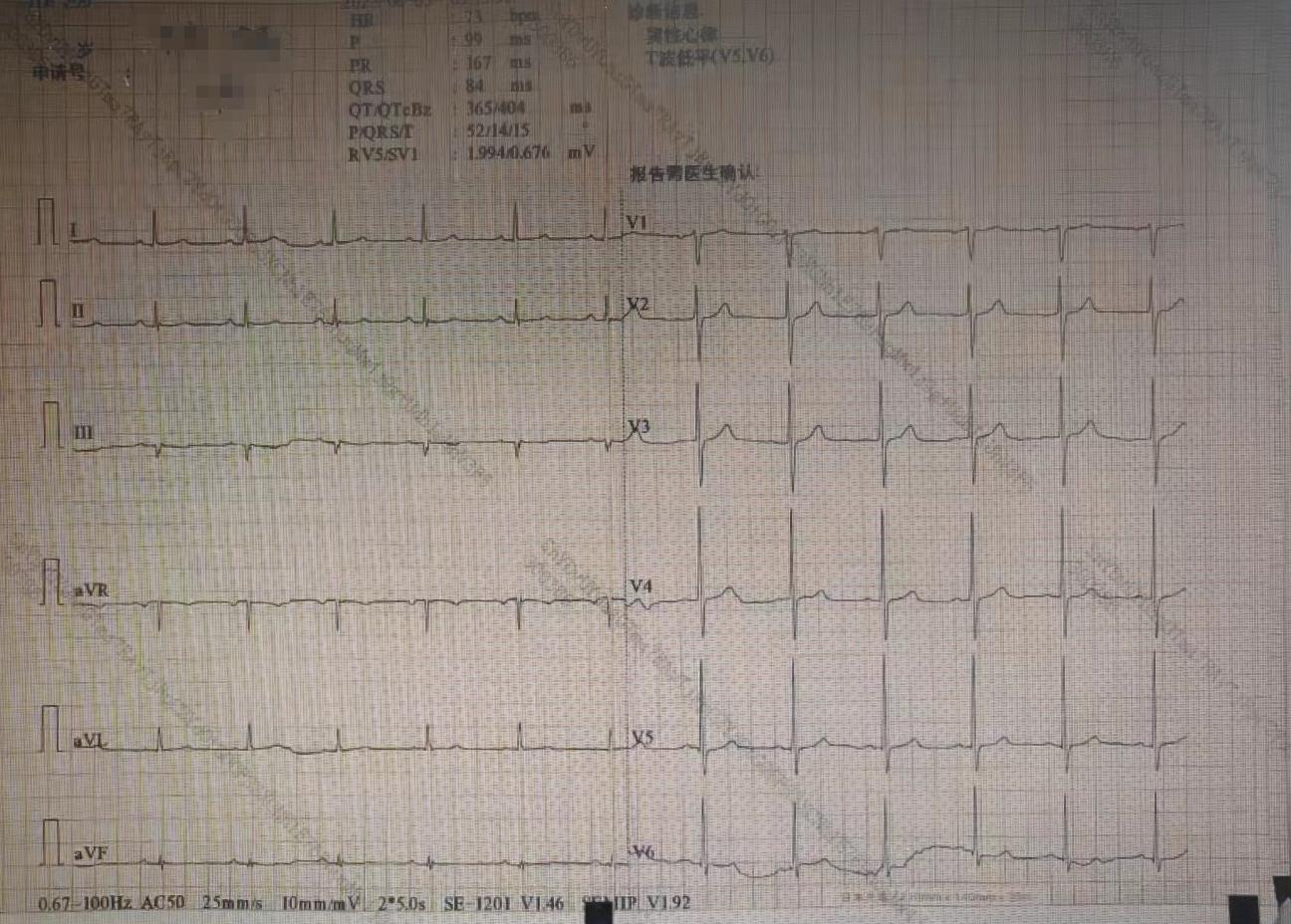
Renal function: BUN 6.00mmol/L,Cr: 73.7ummol/L, Glomerular Filtration Rate: 86.7ml/min, Blood glucose:6.26mmol/L, LDL:1.39mmol/L, CK-MB <2.0 ng/ml, Troponin I <0.010 ng/mlWhole blood count: Normal, Echocardiogram: LV size normal, EF: 66%, ECG: Normal Sinus rhythm.
Relevant Catheterization Findings
LAD: severe calcification, proximal LAD with stenosis of about 90%, LCX: stenosis of about 70-80%, RCA severe calcification, subtotal occlusion at the proximal segment; LAD to RCA distal collateral.
 IM000009.avi
 IM000010.avi
Interventional Management
Procedural Step
We used 7French AL 0.75 (Cordis) guiding to engage the RCA. And then implanted temporary pacemake lead (5F Medtronic) to the apic of right ventricle. We sent one Sionblue 0.014" guidewire (ASAHI) to the distal of the RCA, and then used Corsair Microcatheter (ASAHI) to change Rotawire drive floppy (Bostonscientific) to the distal of the RCA. Used 1.25 mm Burr (Bostonscientific) to do atherectomy with 160 -180 K/minute from proximal to the distal of RCA. And examed the lesion by intravascular ultrasound (IVUS, Bostonscientific) found there were 360 degree calcification lesions, minimum lumen area (MLA) 3.23 mm square. Sent 3.0 x12 mm Intravascular Lithotripsy (IVL) balloon to the distal of RCA by the support with 6 French Guidezilla (Bostonscientific). Used 60 pulses of IVL from distal to the proximal of RCA. And then implant distal stent by 2.5 x 32mm (Bostonscientific, Promus PREMIER), middle stent 3.0 x 32mm (Bostonscientific, Promus PREMIER), and 3.5 x 28mm stent (Bostonscientific, Promus PREMIER) at proximal RCA. Post dilated the stents by 2.75 x 12mm, 3.0 x 12mm, 3.5 x 12mm, 4.0 x 12mm NC balloon (Bostonscientific, NC EMERGE). Repeat IVUS exam and final angiogram shown a good result.
 IM000017.avi
 IM000035.avi
 IM000058.avi
Case Summary
Intravascula lithotripsy (IVL) has a low learning curve and is easy to master; It is safer, shorten procedure time and more efficient than rotablation teachnique. It is effective for both superficial and deep calcifications. IVL make impossible lesions to do PCI become to possible. Sometimes, in very complex case, we need to combine rotablation and IVL to get good result.
