CASE20230820_001
The Invisible Seal to Coronary Perforation
By Yuen Fung Yiu
Presenter
Yuen Fung Yiu
Authors
Yuen Fung Yiu1
Affiliation
Princess Margaret Hospital, Hong Kong, China1,
View Study Report
CASE20230820_001
Complication Management - Complication Management
The Invisible Seal to Coronary Perforation
Yuen Fung Yiu1
Princess Margaret Hospital, Hong Kong, China1,
Clinical Information
Relevant Clinical History and Physical Exam
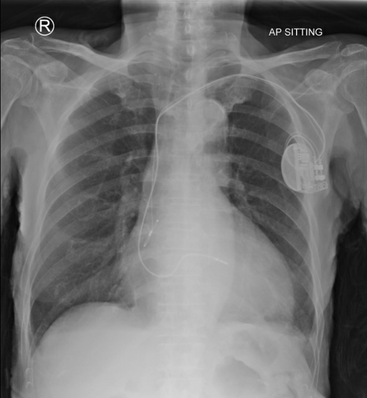
Mr. LC was a 91-year-old chronic smoker. He had a history of hypertension, hyperlipidaemia, impaired fasting glucose and sick sinus syndrome for which he had a pacemaker implanted in 2016. Despite his age, he was capable of performing activities of daily living without assistance.
Relevant Test Results Prior to Catheterization
Serum creatinine: 114 μmol/L
Relevant Catheterization Findings
LM: normal
 1. Diagnostic shot.avi
Interventional Management
Procedural Step
PCI was done via the right femoral artery as the right radial pulse was absent. LCA was engaged with a 6Fr EBU 3.75 guide catheter.
2. Post-stenting perforation.avi
3. Delivery of suture.avi
4. Final.avi
Case Summary
Guidewire induced-distal coronary artery perforation is relatively uncommon, but possessing the knowledge to effectively manage it is of utmost importance. Embolization with absorbable suture is a rapid and inexpensive method. The invisibility of the suture material represents both advantage and disadvantages within this technique.