CASE20230816_001
Angiography Derived 3D-physiologic Mapping of the Coronary Flow Using QFR for the Planning of Complex PCI
By Oyunkhand Buyankhishig, Gereltuya Choijiljav, Bum-Erdene Batbayar, Damdinjav Enkhee, Batzaya Tsognemekh, Erdembileg Dandar, Surenjav Chimed, Batmyagmar Khuyag
Presenter
Oyunkhand Buyankhishig
Authors
Oyunkhand Buyankhishig1, Gereltuya Choijiljav1, Bum-Erdene Batbayar2, Damdinjav Enkhee2, Batzaya Tsognemekh3, Erdembileg Dandar4, Surenjav Chimed5, Batmyagmar Khuyag1
Affiliation
Intermed Hospital, Mongolia1, First State Central Hospital, Mongolia2, National Cardiovascular Center In Mongolia, Mongolia3, Mongolian Interventional Cardiology Research Center, Mongolia4, Third State Central Hospital, Mongolia5,
View Study Report
CASE20230816_001
Complex PCI - Multi-Vessel Disease
Angiography Derived 3D-physiologic Mapping of the Coronary Flow Using QFR for the Planning of Complex PCI
Oyunkhand Buyankhishig1, Gereltuya Choijiljav1, Bum-Erdene Batbayar2, Damdinjav Enkhee2, Batzaya Tsognemekh3, Erdembileg Dandar4, Surenjav Chimed5, Batmyagmar Khuyag1
Intermed Hospital, Mongolia1, First State Central Hospital, Mongolia2, National Cardiovascular Center In Mongolia, Mongolia3, Mongolian Interventional Cardiology Research Center, Mongolia4, Third State Central Hospital, Mongolia5,
Clinical Information
Relevant Clinical History and Physical Exam
A 62 old male patient who had persistent angina regardless of optimal medical treatment. Multiple cardiovascular risk factors including smoking, hypertension and hypercholestrolemia. Prior medications are antihypertensives and aspirin.


Relevant Test Results Prior to Catheterization
Relevant Catheterization Findings
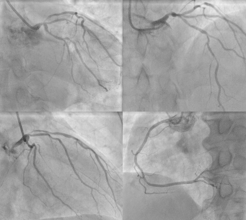
Diagnostic CAG showed diffuse lesions in LM and LAD including severe stenosis in distal LM and moderate stenosis in mid-LAD. LCx and RCA were normal and no significant stenosis.
Interventional Management
Procedural Step
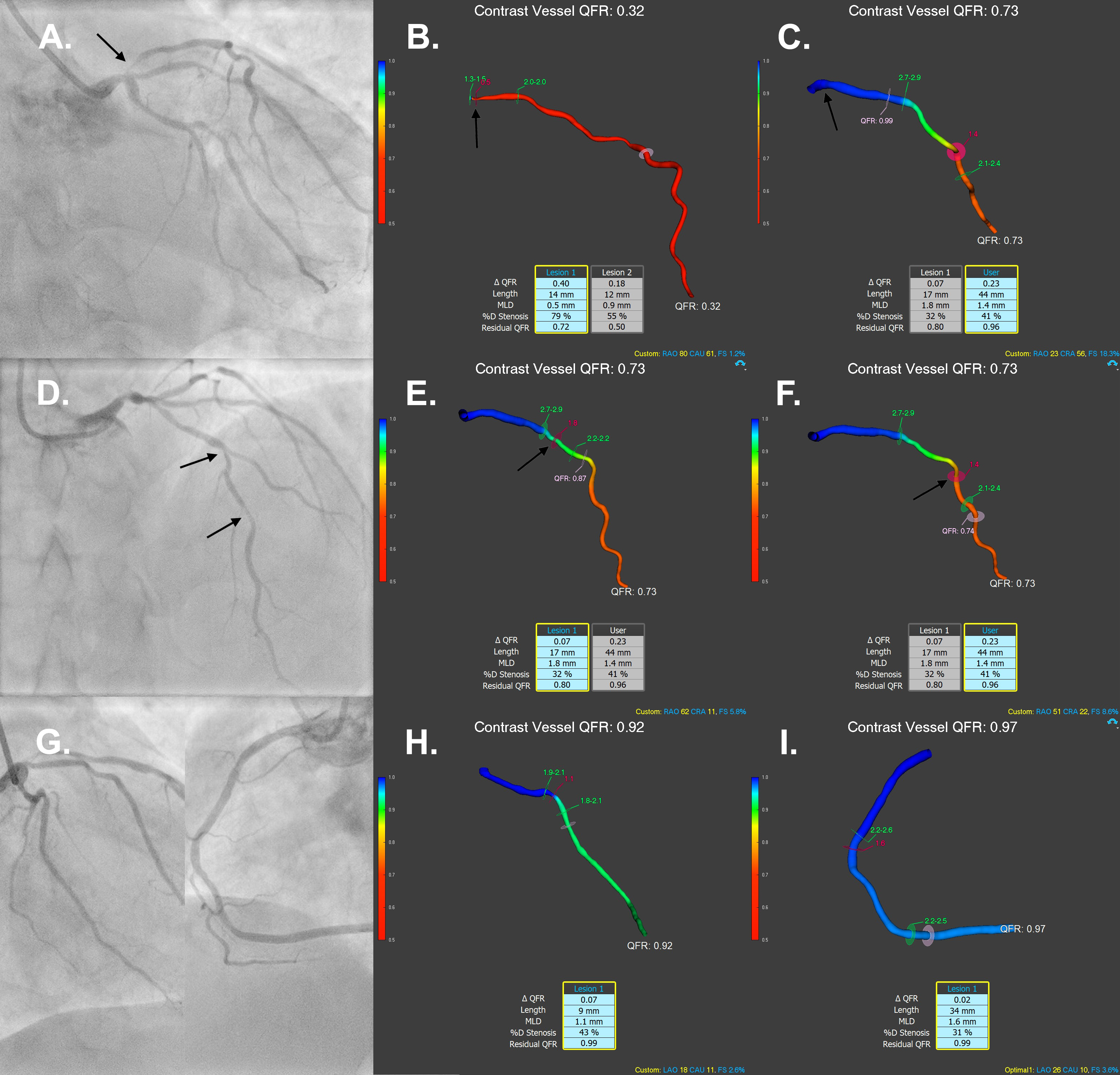
Ondiagnostic angiography, there was critical stenosis at the ostial LAD withthe vessel QFR of 0.32 (arrow, Figure 1A and 1B). The proximal LAD lesion wassuccessfully treated with drug-eluting stent (DES) which was implanted from LMSto proximal LAD with final post-stenotic QFR of 0.99 (arrow, Figure 1C). Therewere another two isolated lesions (arrow, Figure 1D) with a total length of44mm in the mid LAD which have had post-stenotic QFR of 0.87 and 0.74,respectively (arrow, Figure 1E and 1F). The mid LAD lesions were untreated atthe moment because vessel QFR were borderline and the patient was left onoptimal medical treatment. Staged PCI could be considered for mid LAD lesionsif the patient is symptomatic despite optimal medical treatment. The leftcircumflex (LCx) and right coronary artery (RCA) were normal (Figure 1G, 1H and1I).


Case Summary
Theangiography image based 3D-physiologic mapping of coronary physiologyusing the QFR could be a cost effective and non-invasive alternative topressure wire based invasive approaches to guide complex PCI.
