CASE20210827_003
Use of iFR in Coronary Spasm Case of a Pre-Transplant CKD Patient: To Stent or Not to Stent
By , ,
Presenter
Indah Sukmawati
Authors
1, 1, 1
Affiliation
, Indonesia1
Imaging - Physiologic Lesion Assessment
Use of iFR in Coronary Spasm Case of a Pre-Transplant CKD Patient: To Stent or Not to Stent
1, 1, 1
, Indonesia1
Clinical Information
Patient initials or Identifier Number
AR
Relevant Clinical History and Physical Exam
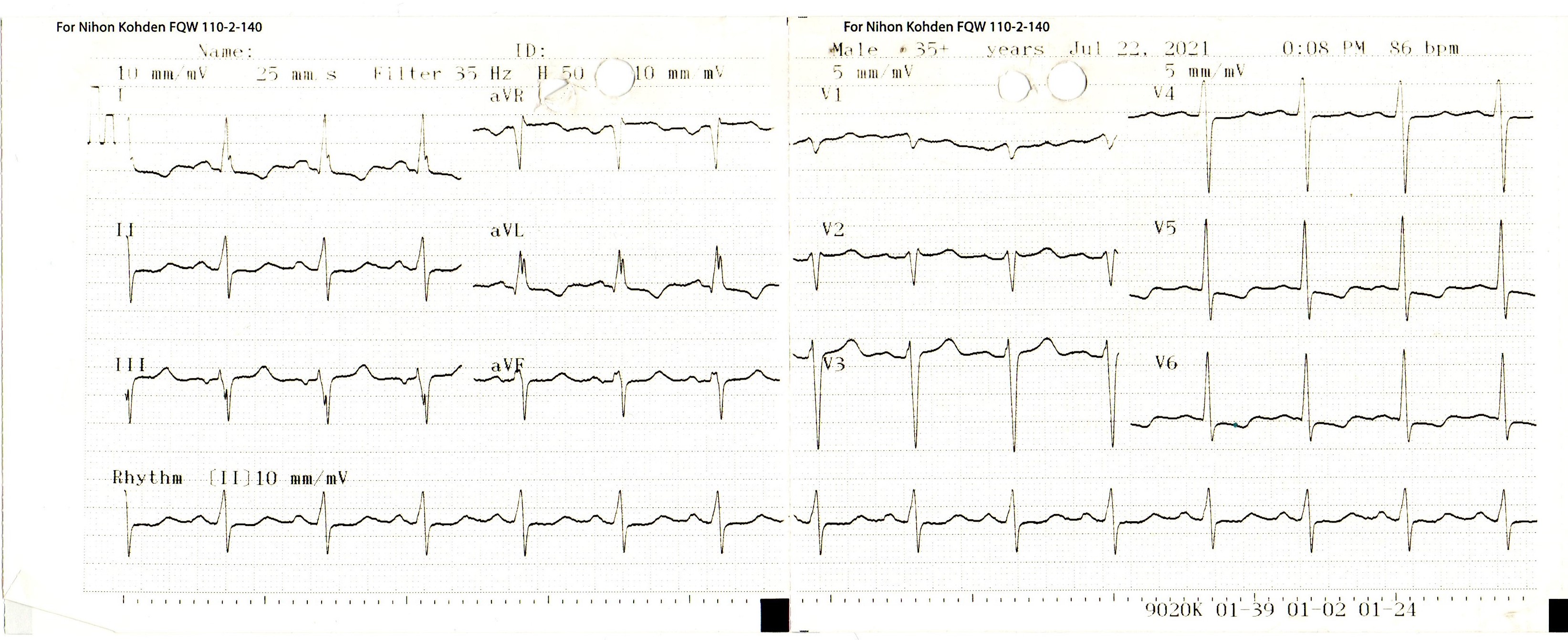
A 45 year-old-male patient was referred for coronary evaluation prior to kidney transplant. He was asymptomatic with history of diabetes, hypertension and kidney failure on routine dialysis. He had a high blood pressure of 197 / 88 mmHg and apical systolic murmur and AV fistula (Cimino) on his left arm. His other examination was unremarkable. He was already on aspirin 80 mg, rosuvastatin 20 mg and fixed-dose hypertension medication amlodipin/valsartan 5 / 80 mg.
Relevant Test Results Prior to Catheterization

Relevant Catheterization Findings
Coronary angiogram showed normal right coronary artery, left main and left circumflex. Mid LAD had 90 % stenosis and distal LAD had 80 % stenosis which both were suspected to have caused by coronary spasm. Post intracoronary nitroglycerin injection, the angiogram showed borderline stenosis of 49 % and 60 % in mid and distal LAD, respectively.
 RCA Angiogram.wmv
 LCA angiogram - spasm.wmv
 LCA Post Intracoronary Nitroglycerin.wmv
Interventional Management
Procedural Step
To confirm whether the borderline stenosis limit the flow, an iFR examination was performed. A Verrata pressure wire was inserted through a 6F system with RU guiding catheter to LAD. Significant pressure gradients were observed with spot iFR of 0.84 and 0.79 in mid and distal LAD, respectively. Pullback iFR was 0.76 in LAD. Direct stenting PCI to mid and distal LAD was then decided.
 3. Final LAD Angiogram.mp4
 1. iFR.mp4
 2. PCI distal and mid LAD with 2 DES.mp4
Case Summary
In a case of significant stenosis caused by coronary spasm with borderline stenosis on angiogram after intracoronary nitroglycerin injection, iFR was useful in evaluating the pressure gradient which in this case showed flow-limiting stenosis thus prompting necessary PCI to be done.
