
CASE20210816_005
OCT Guided PCI for Restenotic Lesions- A Case Series
By
Presenter
Rohit Mody
Authors
1
Affiliation
, India1
Imaging - Invasive Imaging (IVUS, OCT, spectroscopy, etc)
OCT Guided PCI for Restenotic Lesions- A Case Series
1
, India1
Clinical Information
Patient initials or Identifier Number
Case 1- 154238 Case 2- 196442
Relevant Clinical History and Physical Exam
Case1- 59 year old Female Post-PTCA to LAD A known case of Hypertension Diabetes, Normal LV Functions, AOE 3
Case2- 74 year Old Male Post PTCA- to LAD Known case of Hypertension, AOE 3, non DiabeticEF- 35%
Relevant Test Results Prior to Catheterization
Case 1- Normal LV FunctionsCase 2-EF- 35%
Relevant Catheterization Findings
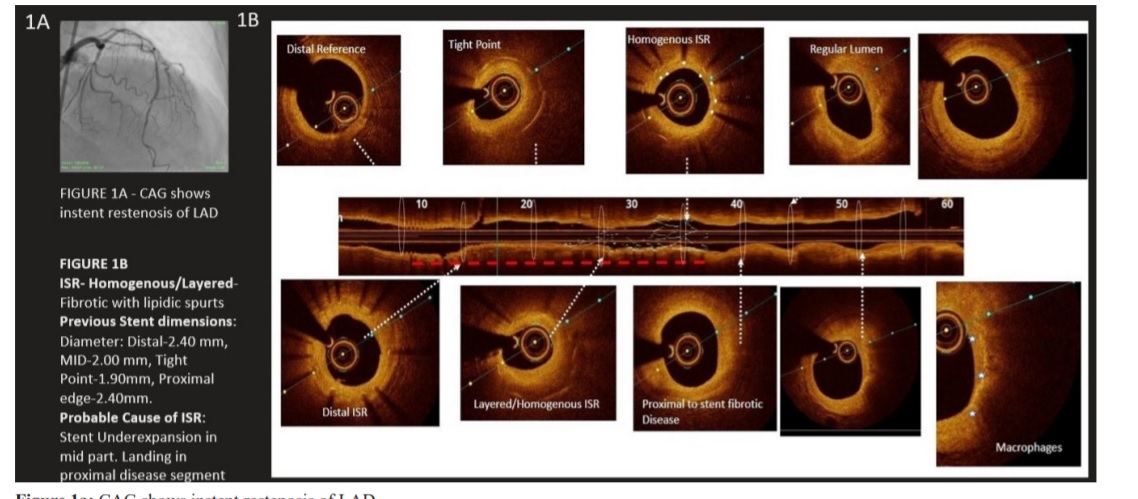
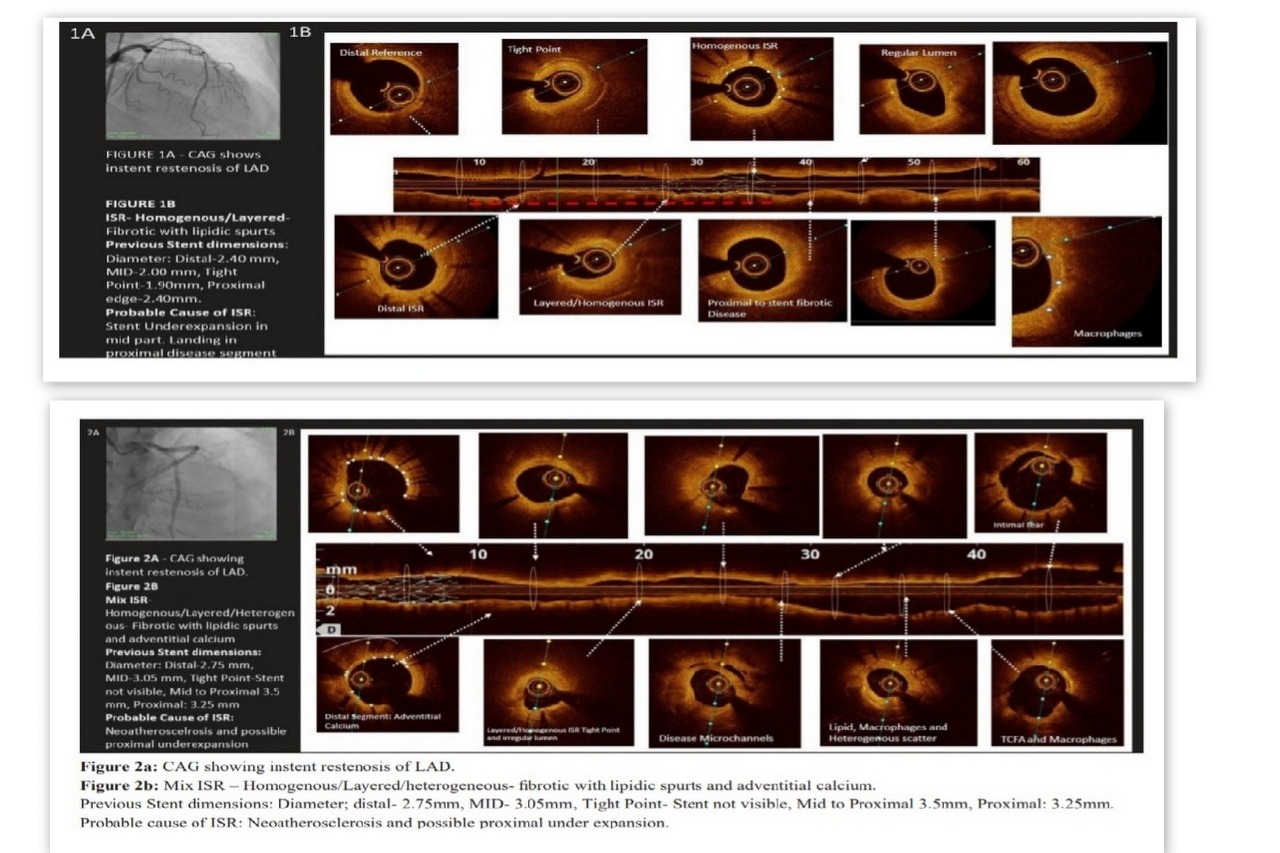
Case1- Angiography reveals A long lesion from Proximal LAD to Proximal edge of restenosis of LAD stent
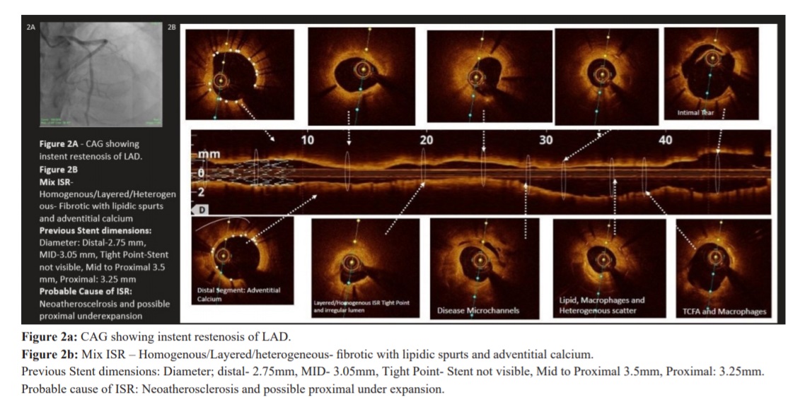
case2- Angiography revelas LAD restenosis
Interventional Management
Procedural Step
Case1-Pre-PTCA OCT Run,Stent Distal 3X34mm deployed at 10 ATM,Post Dilatation with 3.0X15mm Balloon,Stent Deployed 3.5X23mm proximal to mid LAD at 10ATM,OCT Run done. No complications, TIMI 3 flow achieved Good final results
Case2-LAD was crossed with BMW Guide Wire,OCT Done,Pre Dilatation done with 3.5X14mm Balloon at 20 atm,Stent Deployed in Proximal to Mid LAD 3.0X33mm at 10 ATM,Another Stent Deployed 4.0X18mm in LMCA to proximal LAD overlapping Distal Stent at 10 ATM,OCT runs after Post Dilatation. No complications, TIMI 3 flow
Case2-LAD was crossed with BMW Guide Wire,OCT Done,Pre Dilatation done with 3.5X14mm Balloon at 20 atm,Stent Deployed in Proximal to Mid LAD 3.0X33mm at 10 ATM,Another Stent Deployed 4.0X18mm in LMCA to proximal LAD overlapping Distal Stent at 10 ATM,OCT runs after Post Dilatation. No complications, TIMI 3 flow
Case Summary
OCT has the ability to identify differential patterns of restenotic tissue after stentingIt has also generated a hypothesis that with time instent neo atherosclerosis produces unstable characteristics into plaque and that can be possible cause of late stent thrombosis seen in DES patients and this can be characterized by OCT