
CASE20220826_024
Changing the Course of Action through Imaging
By ,
Presenter
Su Hnin Hlaing
Authors
1, 1
Affiliation
, Australia1
Imaging - Invasive Imaging (IVUS, OCT, spectroscopy, etc)
Changing the Course of Action through Imaging
1, 1
, Australia1
Clinical Information
Patient initials or Identifier Number
VW
Relevant Clinical History and Physical Exam
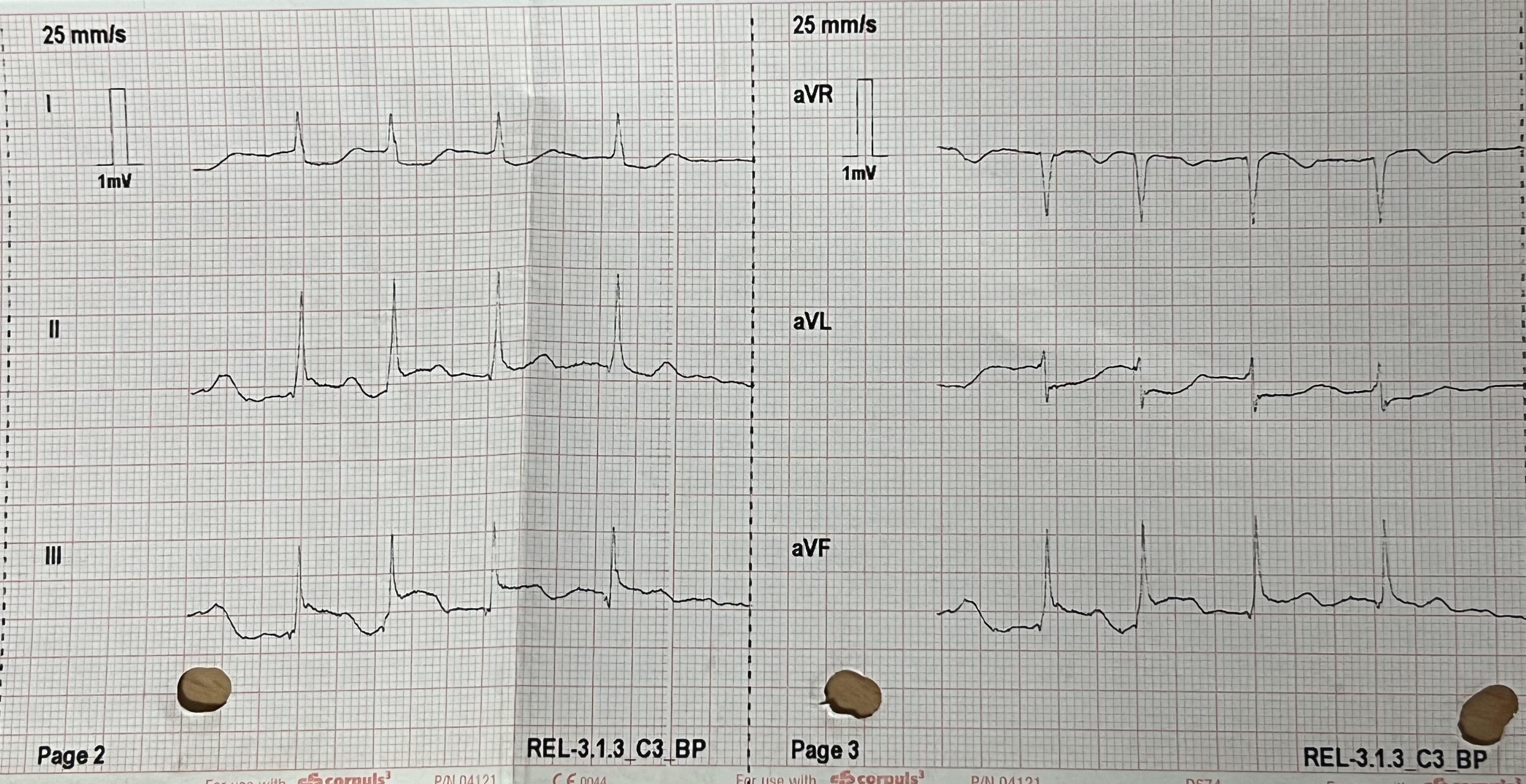
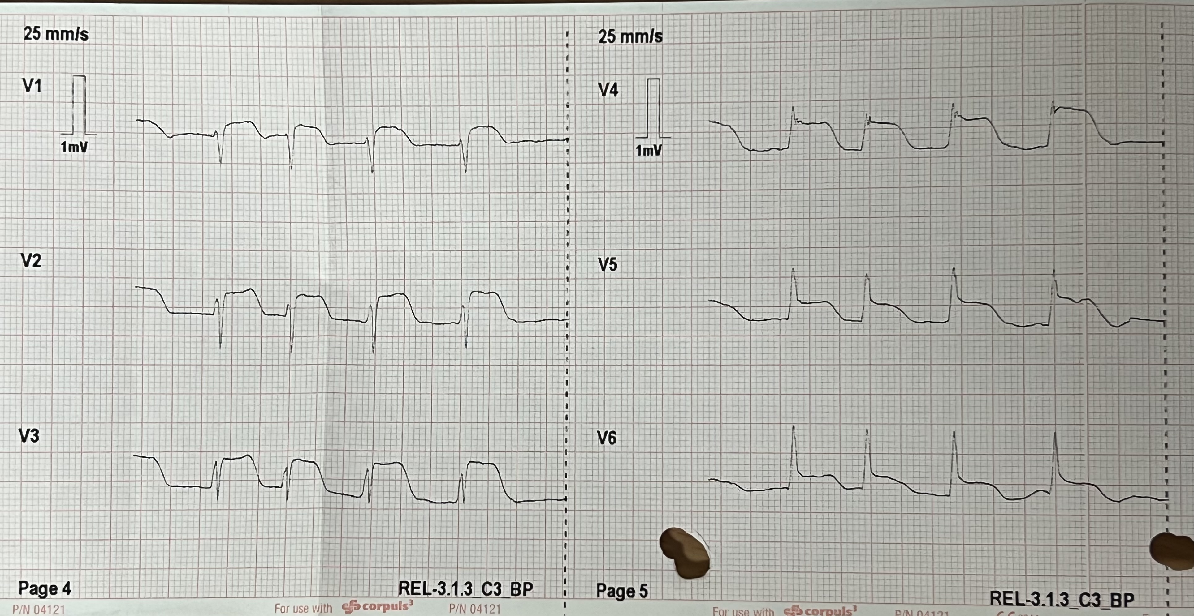
o63 year old female presented to ED at 15:37 with burning epigastric pain since morning. Background: Hypertension, Hypercholesterolemia, COPD- current smoker , High BMI 41.5, CT calcium score 683 and ESE in 2017 - no exercised-induced ischemia at low workload Abdomen - soft, minimal tenderness in epigastrium on palpationInitial impression was likely gastritis and discharged at 21:30 with outpatient MPS. Ongoing pain and called ambulance at 00:19 and ECG showed widespread ST elevation MI
Relevant Test Results Prior to Catheterization
1st troponin at 17:30 - 7ng/L, 2nd troponin at 19:43 8ng/L Troponin at 1:35 - 36,405 ng/L
 RCA LAO CRA.avi
LCA RAO CRA.avi
LCA RAU CAU.avi
Relevant Catheterization Findings
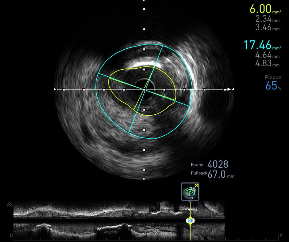
There was no significant disease in RCA. Severe stenosis in l eft main and proximal LAD. IVUS showed long dissection plane and haematoma from left main to distal LAD with areas of severe stenosis in left main and proximal LAD.
IVUS LAD..avi
TOE 1.mpg
TOE color.mpg
Interventional Management
Procedural Step
Patient was in cardiogenic shock. IABP was inserted. After discussion with cardiothoracic surgeon, decided for emergency CABG. Bedside TTE showed evidence of dilated aortic root with eccentric AR which was not changed significantly from previous TTE done in 2021. Normal LV systolic function 64% TEE done in OT showed Type A dissection, dissected proximal LAD (long segment) and intimal tear noted in ascending aorta. Ascending aorta replacement with 28mm Intergard straight graft and aortic valve repair and CABG x 2 - SVG - LAD and SVG D1 was performed by CTS Patient was discharged home after 2 months stay in hospital
with wires.avi
Case Summary
IVUS plays a pivotal role in not only providing detailed information on the location and extent of dissection but also directing the guide wire into the true lumenIVUS has changed the course of the action for this patient Transoesophagel echo is one of the important diagnostic modalities in patients with acute aortic dissection